Drug Self-Administration in Head-Fixed Mice
Of the preclinical models, drug self-administration has the greatest construct and predictive validity and its use has lead to groundbreaking discoveries in addiction research. This approach capitalizes on how animals, much like humans, will choose to seek drugs of abuse of their own volition and, much like humans, relapse in response to the same general stimuli.
However, while this technique is foundational in addiction research, freely moving animal preparations are incompatible with emerging technologies— such as two-photon microscopy and holographic optogenetics— that could allow greater resolution of potential treatment targets.
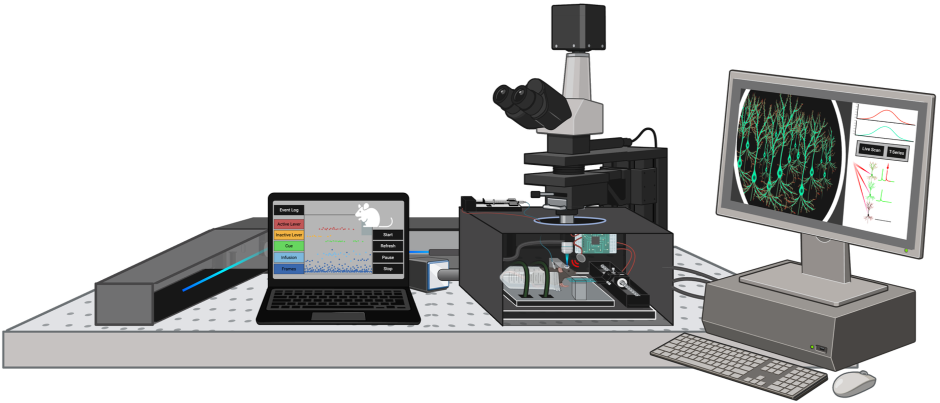
To overcome this, during her postdoctoral Dr. Doncheck and a team of scientists worked together to develop a modified version of drug self-administration wherein mice are head-fixed, allowing for them to be positioned under a two-photon microscope while engaging in drug seeking. Multiple layers of setup are required to implement head-fixed drug self-administration— custom operant equipment is 3D-printed, wired, and operated via open-source software (MatLab legacy and REACHER platform-based versions exist), and head-fixation equipment is generated in collaboration with the UAB Machine Shop Core. Delicate surgeries must be performed to allow head-fixation, in vivo two-photon imaging, and intravenous drug delivery. Niche adaptations and analyses, such as individualized lever placement and discrimination indices, are requisite for successful implementation of behavioral experiments. Please see Doncheck et al., 2026 for a detailed protocol.
Two-Photon Microscopy & Holographic Optogenetics
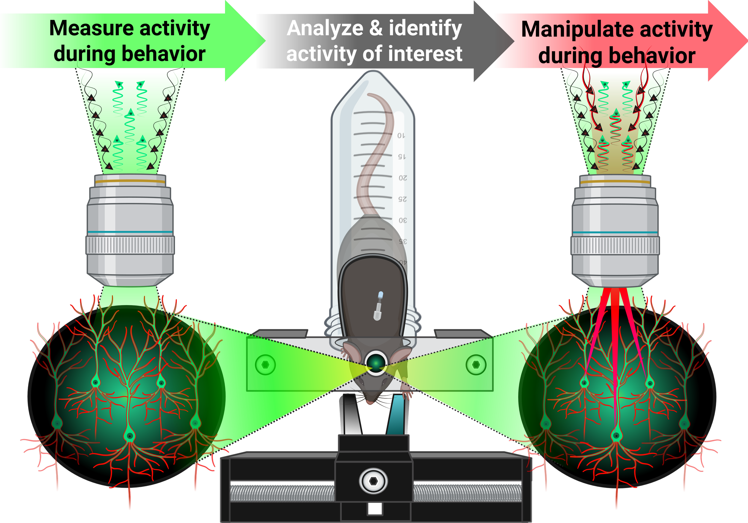
To study neural activity with single-cell, sub-second resolution, the Doncheck Lab uses two-photon microscopy, a powerful approach for capturing rapid changes in cellular activity. Two-photon microscopy is based on near-simultaneous absorption of two lower-energy photons to excite a fluorescent molecule— an event that only occurs at the microscope’s focal plane. This spatial confinement minimizes light scattering, photobleaching, and tissue damage, while use of longer-wavelength light enables deep penetration into intact brain tissue. As a result, this technique is ideally suited for in vivo imaging of individual cells, circuits, and dynamic processes in the living brains. The lab is equipped with a state-of-the-art Bruker Ultima 2Pplus system and a Coherent Chameleon Discovery NX laser with dual outputs.
The system is also equipped with a second, powerful fixed-wavelength laser (Coherent Monaco) and NeuraLight 3D Ultra liquid crystal spatial light modulator, which alters light phase wavefronts to create computer-generated holograms for simultaneous single-cell photostimulation during imaging.
Whole-Cell Patch-Clamp Electrophysiology
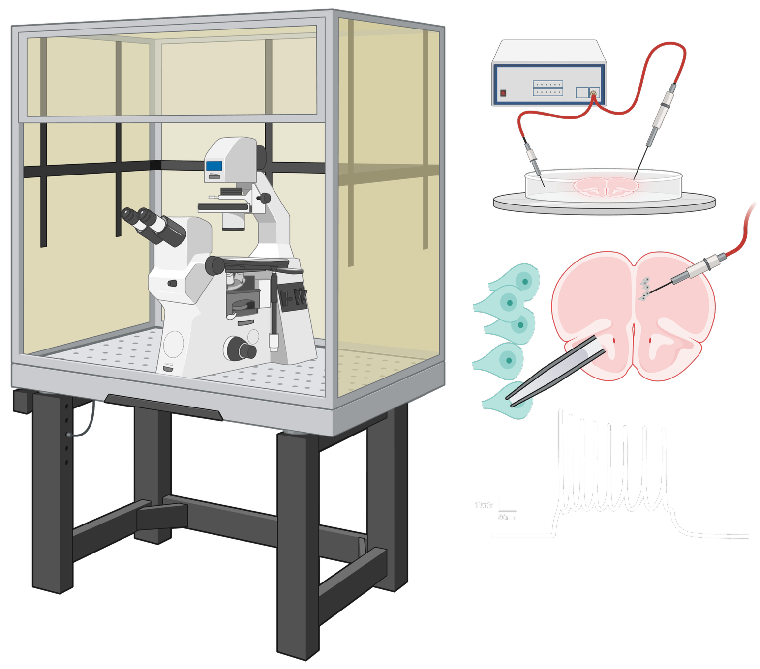
Whole-cell patch-clamp electrophysiology allows for direct measurement of the activity of individual cells. Using a fine glass recording electrode, we can assess how neurons respond to neurotransmitters, hormones, and other experimental manipulations, providing insight into the cellular mechanisms that regulate neural function.
In our laboratory, we use whole-cell patch-clamp recordings to investigate how neuroendocrine signaling shapes neuronal excitability and synaptic transmission within circuits involved in motivated behavior. This approach enables us to identify cellular and circuit mechanisms that contribute to sex differences in brain function and behavior, and to better understand how endocrine and neural signals interact to influence behavior in both health and disease states.
Hormone Measurement & Phase Monitoring
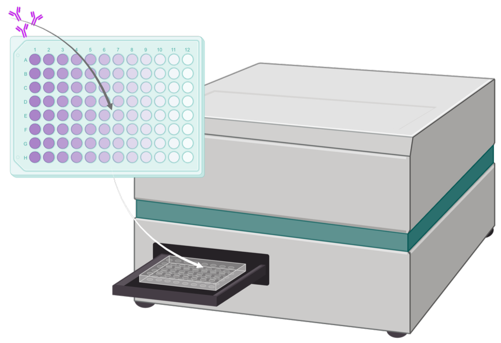
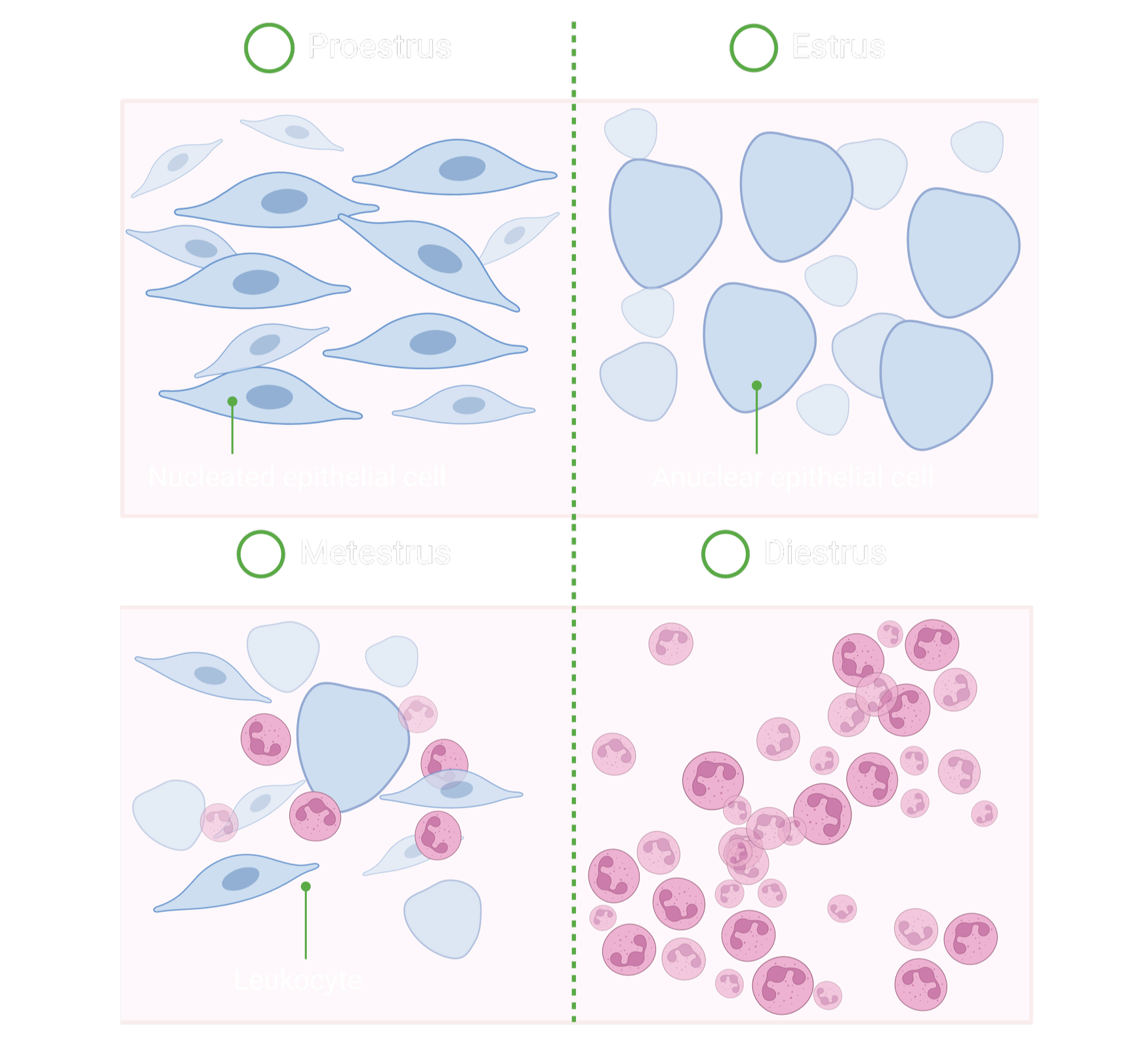
As hormones can profoundly influence neural activity and behavior, our laboratory incorporates hormone measurements and estrous cycle monitoring into studies of drug seeking and neural function. Enzyme-linked immunosorbent assays (ELISAs) are used to quantify circulating hormone levels, providing a sensitive and reliable measure of endocrine state at the time of behavioral testing or neural recordings. To determine estrous cycle stage, we collect vaginal lavage samples and assess cellular composition using Papanicolaou staining. This approach allows identification of the stages of the estrous cycle based on characteristic changes in cell populations, such as morphology or nucleated appearance of epithelial cells and the presence of leukocytes.
By combining hormone measurements with estrous cycle monitoring, we can evaluate how fluctuations in ovarian hormones influence drug-seeking behaviors, neural activity, and neural circuit function. These methods provide an important framework for understanding the biological mechanisms underlying sex differences in addiction and other motivated behaviors.
Behavioral Pharmacology & Chemogenetics
Behavioral pharmacology and chemogenetics are central components of our research program and provide complementary approaches for investigating the neurobiological mechanisms underlying addiction-related behaviors. By combining pharmacological manipulations with behavioral models of drug seeking, we examine how hormones, neuromodulators, and specific receptor signaling systems influence motivated behavior. In parallel, chemogenetic approaches allow us to selectively manipulate the activity of defined neuronal populations and circuits during behavior. Together, these techniques enable us to establish causal links between neuroendocrine signaling, neural circuit activity, and behavioral outcomes.